

In short
- 01First point of contactOral diagnosis and radiology is usually where a dental visit starts, using examination and X-rays to work out what is wrong.
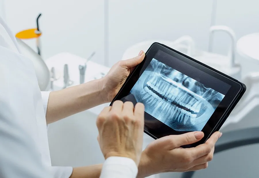
- 02Two imaging typesConventional radiography includes panoramic and bite-wing X-rays, while digital radiography uses less radiation for a similar image.
- 03Full medical pictureYour dentist uses your medical history alongside the exam and X-rays to reach a diagnosis and treatment plan.
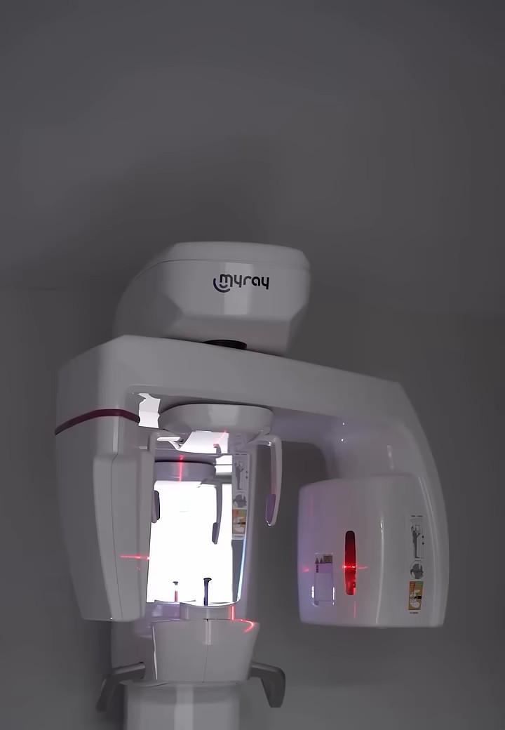
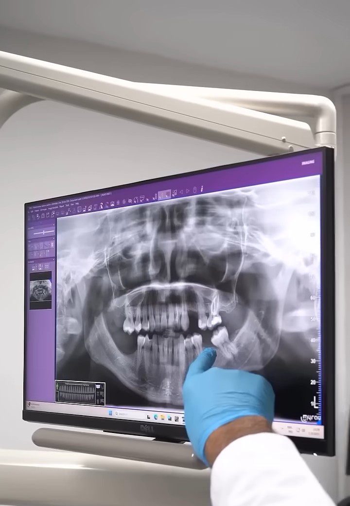
Oral diagnosis and radiology covers the examinations used in dentistry to diagnose problems in the mouth and plan treatment for them. It is usually the first stop when something feels wrong, similar to a pre-examination, and includes procedures such as panoramic X-rays. It looks beyond the teeth and gums to the tissues of the jawbone, and can sometimes pick up signs of conditions elsewhere in the body that show up in the mouth.
What is oral and dental radiology?
Oral and dental radiology uses imaging to help diagnose and treat oral and dental disease, producing images of the structures inside the mouth and teeth that support early diagnosis and treatment. It is used to find:
- Tooth decay
- Gum disease
- Tooth abscess
- Bone fractures
- Tumours
Tooth decay is the most common of these, and treating it early is usually simpler. Gum disease is also common, and early treatment can help prevent tooth loss.
What radiographic methods are used?
Conventional radiography
Conventional radiography shows the mouth and teeth in a single plane. It includes panoramic, periapical, bite-wing and occlusal radiography.
Digital radiography
Digital radiography also images the mouth and teeth, but uses less radiation and produces a higher-quality image than conventional radiography.
How is an oral diagnosis made?
A good oral diagnosis starts with a careful oral and dental examination, covering the teeth, gums, the tissue between them, and soft tissue such as the tongue and palate. Radiological methods are used where the examination findings call for them, with radiation exposure kept to a minimum. The dentist combines these findings, together with your medical history, to reach a diagnosis and build a treatment plan.
With any surgical or interventional procedure, results can vary from person to person.
You are reading about Oral diagnosis and radiology. Ask me anything about it, or pick a question below.
Frequently asked questions
Are X-rays used in oral diagnosis and radiology harmful?
The radiation dose from a panoramic dental X-ray is comparable to about 3 hours in front of a computer, a 1-hour phone call, a 4-hour flight, or two hours of sunbathing. A lead neck collar, vest and apron are worn during the X-ray to protect the rest of the body from unnecessary exposure.
Can radiography be performed during pregnancy?
The radiation used in dental X-rays is very low and is not considered a health risk, but X-rays are generally avoided during pregnancy, or suspected pregnancy, unless there is an urgent medical reason for one. If an X-ray is necessary, a lead apron is worn.
What should I tell my dentist before an oral diagnosis and radiology appointment?
Be open with your dentist and answer every question they ask, including about your general health, not just your teeth. A detail that seems minor to you can matter to the diagnosis.
Videos
Send us your panoramic X-ray
Our professors reply with a treatment plan.